
Plastic landmark anchoring in zebrafish compass neurons
Article Date: 07 January 2026
Article URL: https://www.nature.com/articles/s41586-025-09888-x
Article Image: Figure 1
Summary
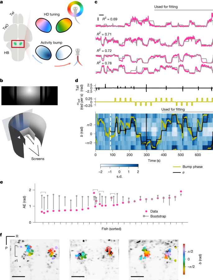
Tanaka and Portugues show that head-direction (HD) neurons in larval zebrafish — gad1b+ cells in anterior hindbrain rhombomere 1 projecting to the dorsal interpeduncular nucleus (dIPN) — can anchor their internal compass to visual landmarks and to rotational optic flow. Using a panoramic virtual-reality projection and two-photon calcium imaging, the authors demonstrate a coherent population ‘bump’ of activity that tracks scene orientation, can be driven by landmark features or optic flow, and remaps plastically after exposure to symmetric (twofold) visual scenes. They identify the left habenula as the primary source of visual landmark input to the dIPN: unilateral ablation of habenula axons abolishes landmark anchoring while leaving optic-flow-based path integration largely intact. The data fit a ring-attractor model with Hebbian visual-to-HD plasticity and reveal candidate angular head velocity (AHV) signals in nearby hindbrain regions.
Key Points
- Larval zebrafish HD neurons form a topographic ring-like map whose population activity is a persistent “bump” that represents heading.
- A panoramic VR (270° × 90°) stimulus shows many HD neurons tune to scene orientation; the bump aligns with visual landmarks in most fish.
- HD cells use both allothetic cues (landmarks via the habenula→dIPN pathway) and idiothetic visual cues (rotational optic flow) to update heading.
- Exposure to symmetric (double-sun) scenes induces plastic remapping: a stretched (2×) mapping from visual space onto the HD array rather than a simple flip–flop.
- Unilateral ablation of visual habenula axons disrupts landmark anchoring but preserves optic-flow-driven integration; putative AHV cells lie in r1–r3 hindbrain clusters.
Content summary
Researchers recorded GCaMP signals from gad1b+ HD neurons in 6–9 dpf zebrafish larvae while projecting panoramic virtual scenes. A sinusoidal fit identified scene-orientation-tuned cells; at the population level their activities formed a moving bump whose phase tracked scene orientation. Closed-loop experiments plus exogenous rotations and translating-dot stimuli showed that the same HD neurons integrate the fish’s turns (motor-related) as well as rotational optic flow.
When scenes were switched (sun-and-bars → Stonehenge) many fish maintained consistent bump–scene offsets, indicating flexibility in landmark use. A three-epoch experiment (smooth rotation / abrupt jumps / noise texture) showed that HD neurons exploit both discrete landmark cues (survive jumps) and optic flow (correlate in noise), with optic-flow gain roughly half of ideal on average.
Introducing a twofold-symmetric scene (double sun) during a learning epoch caused plastic changes: individual HD tunings became bimodal and systematically rotated so that the population effectively mapped 180° of visual space onto 360° of HD space (bump phase ≈ 2×scene). A ring-attractor model with Hebbian learning of all-to-all visual→HD connections reproduces these effects.
Physiologically, visually responsive cells with retinotopic-like receptive fields were concentrated in the left dorsal habenula. Unilateral laser ablation of habenula axons entering the IPN selectively reduced bump–scene alignment (landmark anchoring) without abolishing optic-flow-driven integration. Imaging of the hindbrain revealed candidate AHV/rotation cells with visuomotor tuning in rhombomeres 1–3 that could provide multimodal angular velocity signals.
Context and relevance
This work connects models of ring attractor HD networks with an evolutionarily conserved, non-telencephalic visual route for landmark anchoring. The habenula→IPN projection acts like the insect ring-neuron input to the ellipsoid body, offering an ancient mechanism for visual anchoring of heading. The demonstration of plastic, experience-dependent remapping (including the striking stretched 2× mapping under symmetric cues) highlights how sensory statistics shape internal spatial codes. The identification of candidate AHV and rotation-related elements in the hindbrain advances understanding of how motor and visual signals are integrated for path integration in vertebrates.
Why should I read this
If you care about how brains build and keep a sense of direction, this is tidy, clever work. The authors built a neat panoramic VR for tiny zebrafish, showed the same compass neurons use landmarks and optic flow, and nailed down where visual landmarks come in — the habenula. Also: symmetric scenes do weird, systematic remapping, so don’t assume landmark anchoring is fixed. Short: good methods, clear results, and a few surprising twists.
Author style
Punchy: this paper gives you a compact, well-controlled demonstration that vertebrate compass neurons can flexibly anchor to visual landmarks via the habenula, while still integrating self-motion cues. The experiments are mechanistic (ablation, panoramic VR, modelling) and the findings are broadly relevant — evolutionary parallels to insect compasses, a plastic visual→HD mapping, and candidate AHV nodes. Read the full paper if you want the data and model details; it’s a strong piece for anyone studying navigation, neural attractors, or sensory learning.